
In-vivo, In-vitro, and In-silico Models of Severe Mental Disorders
Welcome to my blog! This summer, I am embarking on an exciting outreach adventure by sharing my PhD research with you. Each week, I will release a new blog post where I unpack a specific aspect of my scientific work. The best part? I will be presenting the information in bite-sized, easily understandable chunks of text! So, whether you are a fellow academic, a curious mind, or simply looking to expand your knowledge, this blog is here to serve you. Throughout the text and in each image you can find links to more detailed sources of information for the topics I discuss here. This week, our focus lies on in-vivo, in-vitro, and in-silico models of severe mental disorders. Let's get started!
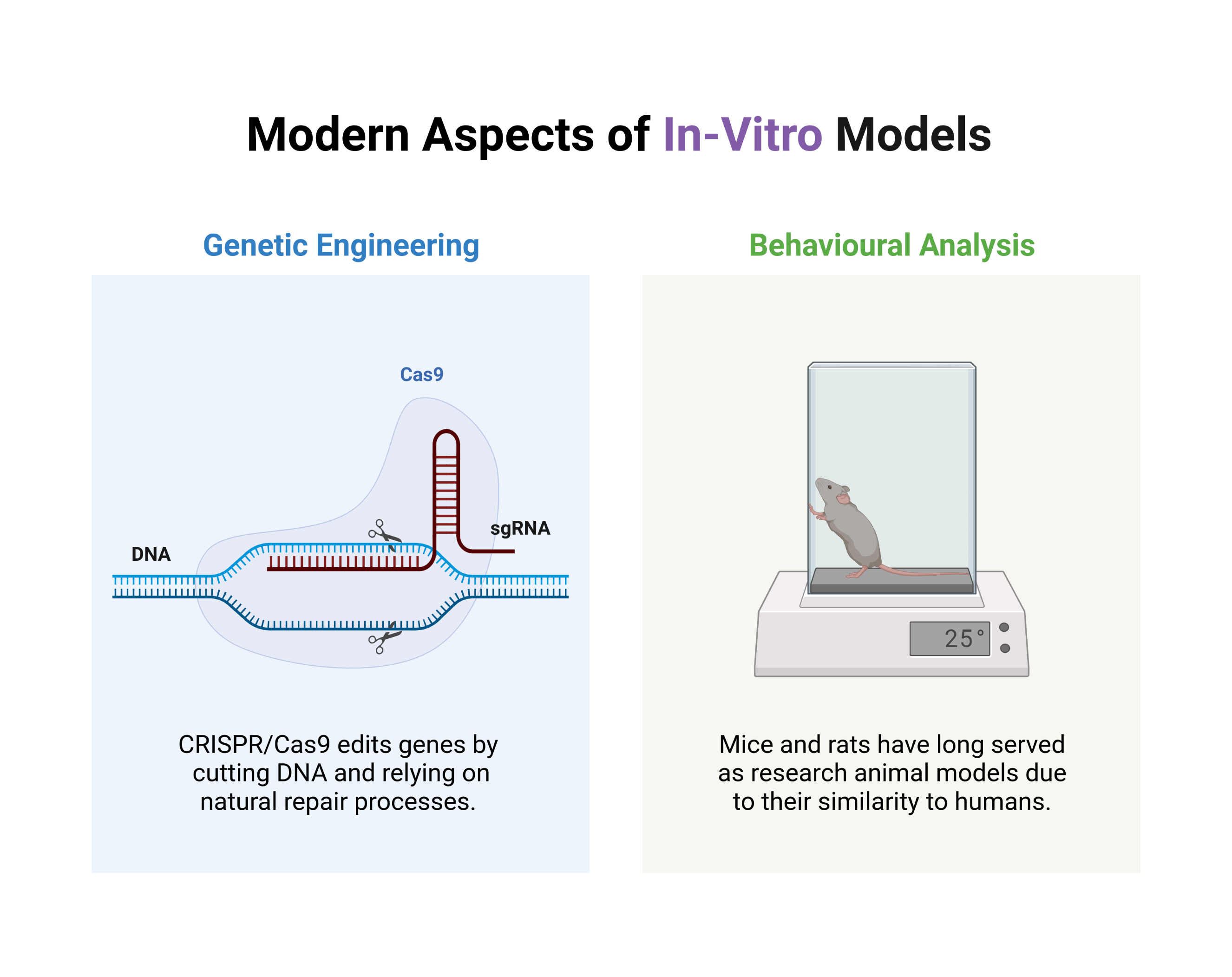
Psychiatric disorders are difficult to study due to their complex genetic architecture, developmental nature, and the lack of direct access to the affected neural tissue of patients. Hence, to validate and interrogate the effect of genetic variations implicated in these diseases, neuroscientists make use of in-vivo, in-vitro, and in-silico models. These disease models (respectively making use of living animals, advanced laboratory techniques, or purely digital means) aim to gain novel insights into a disease or provide the pharmaceutical sector with a strongly supported targets for clinical intervention. After the invention of recombinant DNA technologies in the early 1970’s, genetically altered rodent models started to be used intensely to study a variety of neurological diseases in-vivo. In the case of schizophrenia and bipolar disorder, a significant amount of attention has gone to ion channel gene knock-out rodent models and their aberrant behaviour in working-memory tasks
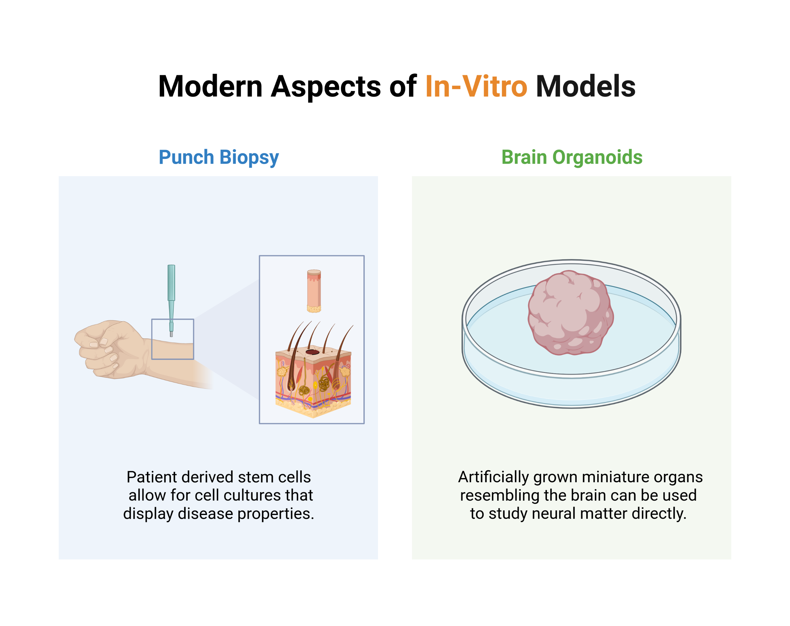
Blood samples (specifically the red blood cells they contain) have been used as a proxy to study protein expression levels in human brain cells. An alternative and significantly more intriguing approach makes use of artificially grown, in vitro, miniature organs resembling the brain, known as brain organoids. These brain organoids are derived from stem cells obtained through skin biopsies, and transformed into a self-organising and three-dimensional cell culture that resembles the cortical structure of the brain, by means of intricate and expensive chemical processes. These miniature brains can be used to study the effects of drugs to screen them for initial safety and efficacy, but they can also be used to compare genetic difference between patients and healthy controls directly in patient-derived neural matter.
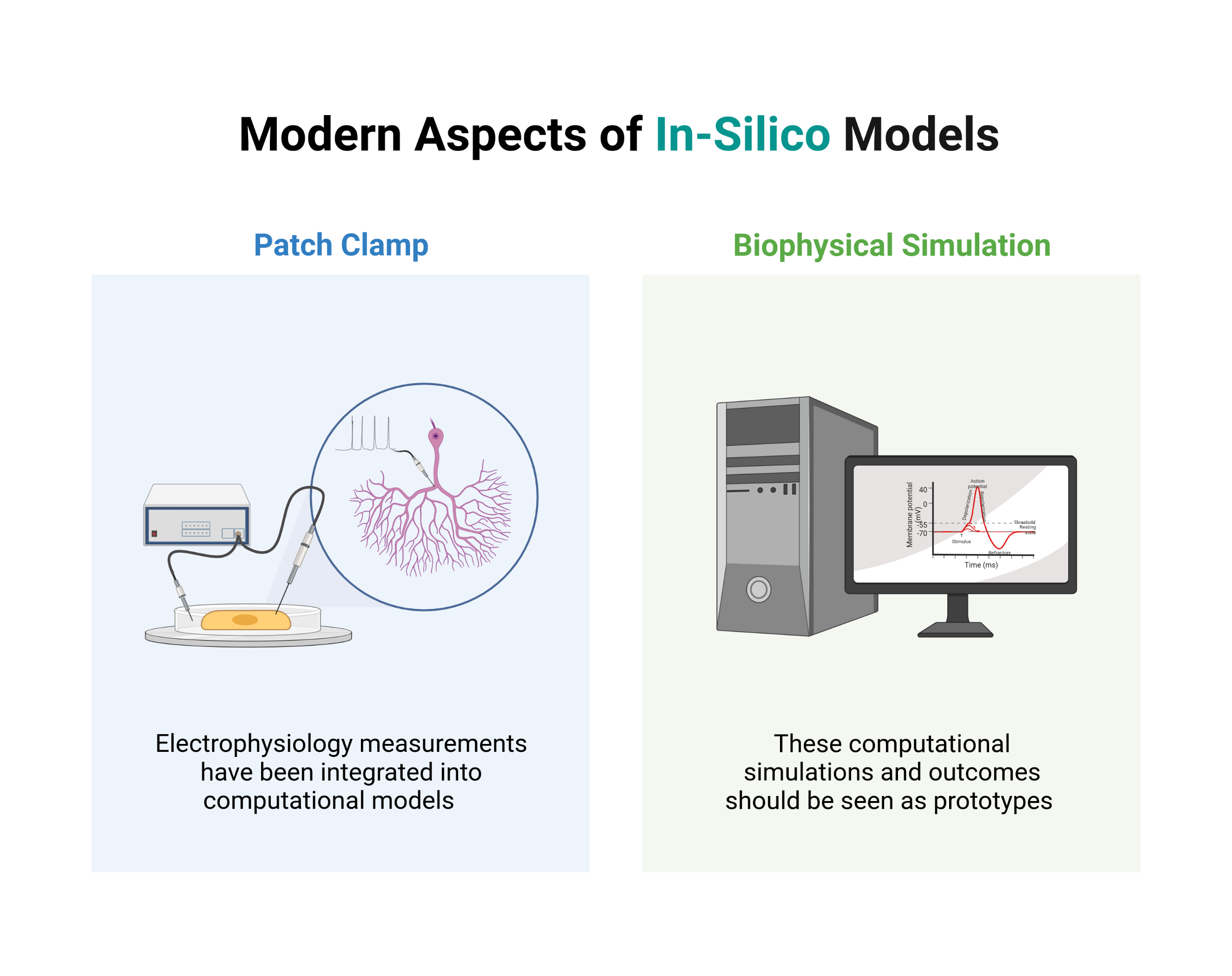
Attempts to evaluate the influence of genetic variants and differentially expressed protein levels through computational models have been made. Due to the challenges in linking genetic mutations to their functional effects, ad-hoc parameter association and modification is often used to link these models to questions regarding the molecular mechanisms of severe mental disorders. Hence, these simulations (and their outcome) should be seen as prototypes rather than being accepted as precise disease models for severe mental disorders. Better knowledge of the functional effect of individual mutations, more accurate biophysically-detailed neuron models, and efficient large-scale biophysically-detailed simulations powered by deep learning approaches are necessary to turn the current methods into tools with real-world applications such as patient stratification or disease mechanism elucidation.
The content of this blog post reflects my personal opinions and insights and should not be attributed to my employer or investors. The information provided in this post is for educational purposes only and should not be construed as medical advice. It is crucial to consult with medical professionals for any mental or physical healthcare concerns. All images featured in this blog post were created using Biorender.com under an academic license. These blog posts are derived from excerpts of my PhD thesis, based on research conducted at the University of Oslo, which you can also read on this website.