
Experimental Advances in Modern Neuroscience
Welcome to my blog! This summer, I am embarking on an exciting outreach adventure by sharing my PhD research with you. Each week, I will release a new blog post where I unpack a specific aspect of my scientific work. The best part? I will be presenting the information in bite-sized, easily understandable chunks of text! So, whether you are a fellow academic, a curious mind, or simply looking to expand your knowledge, this blog is here to serve you. Throughout the text and in each image you can find links to more detailed sources of information for the topics I discuss here. This week's blog post focusses on technological advances in experimental neuroscience. Let's get started!
In our rapidly advancing technological landscape, experimental advances have recently endowed the field of neuroscience with an unprecedented wealth of data. Miniature two-photon microscopes, compact marvels that enable us to simultaneously capture the activity of over a thousand neurons within freely moving lab animals. What is captivating is the intricate choreography of calcium signals they reveal, painting a mesmerizing yet complex, large-scale picture of neuronal interactions. Miniaturized high-density electrical probes, on the other hand, are modern and very delicate tools which are implanted into the brains of lab animals to record the dynamic firing patterns of thousands of individual neurons. This scientific union of these two technological advances allows us to study the firing patterns of individual neurons while simultaneously peering into their collective behavior.
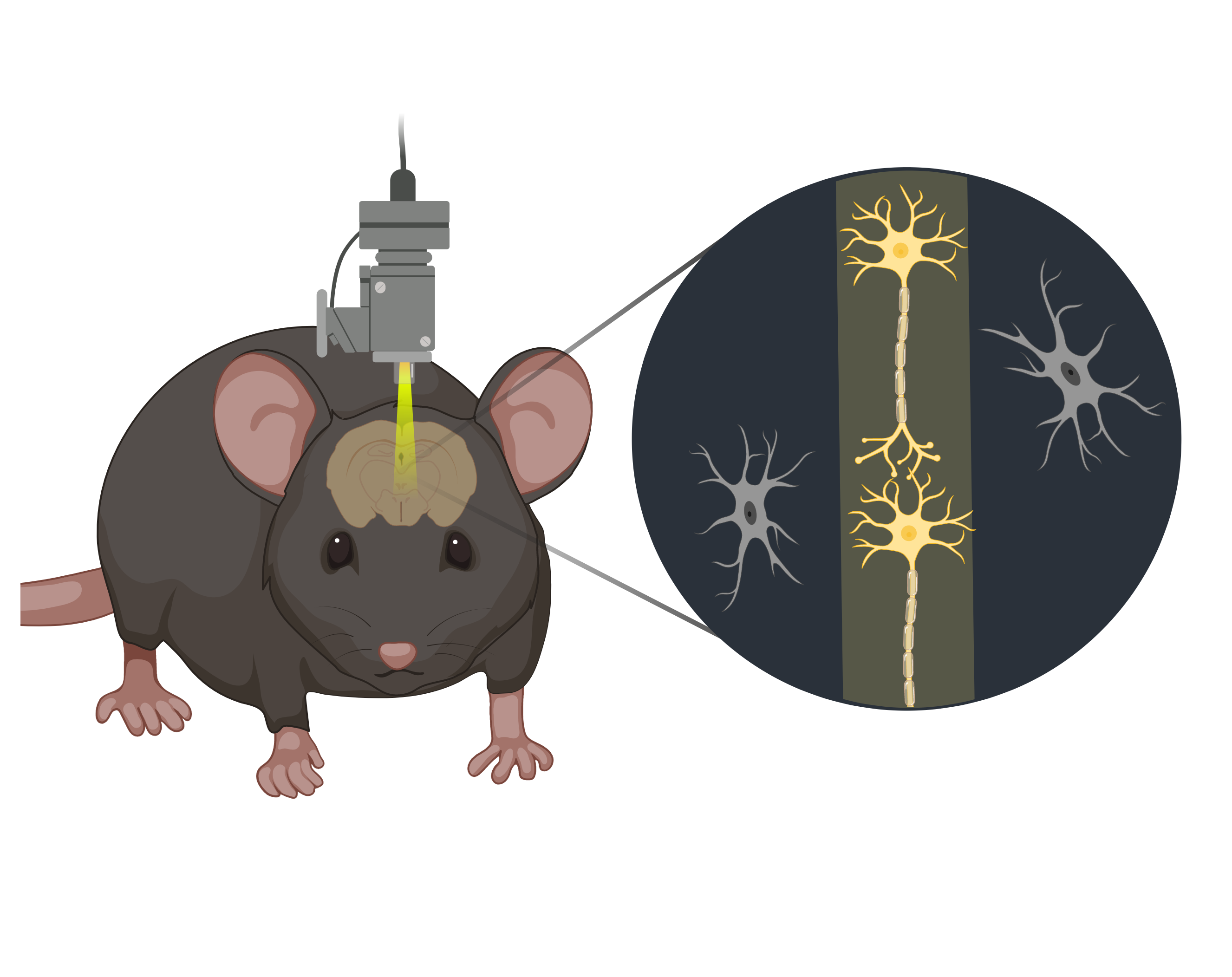
Unlike conventional microscopy methods, two-photon microscopes rely on the simultaneous absorption of two photons to activate fluorescent molecules within the studied specimen. This process enables the visualization of cellular and subcellular structures with unparalleled precision, even within deep layers of living tissue. In the context of neuroscience, two-photon microscopy allows researchers to peer into the dynamic changes in calcium ion concentrations within neurons which play a crucial role in neuronal activity, surging when a neuron fires an action potential. Miniaturized microscopy's significance lies in its ability to provide a minimally-invasive window into the brain's inner workings while animals go about their daily activities, allowing for complex behavioral experiments and extended observations of structural and functional brain plasticity over weeks.
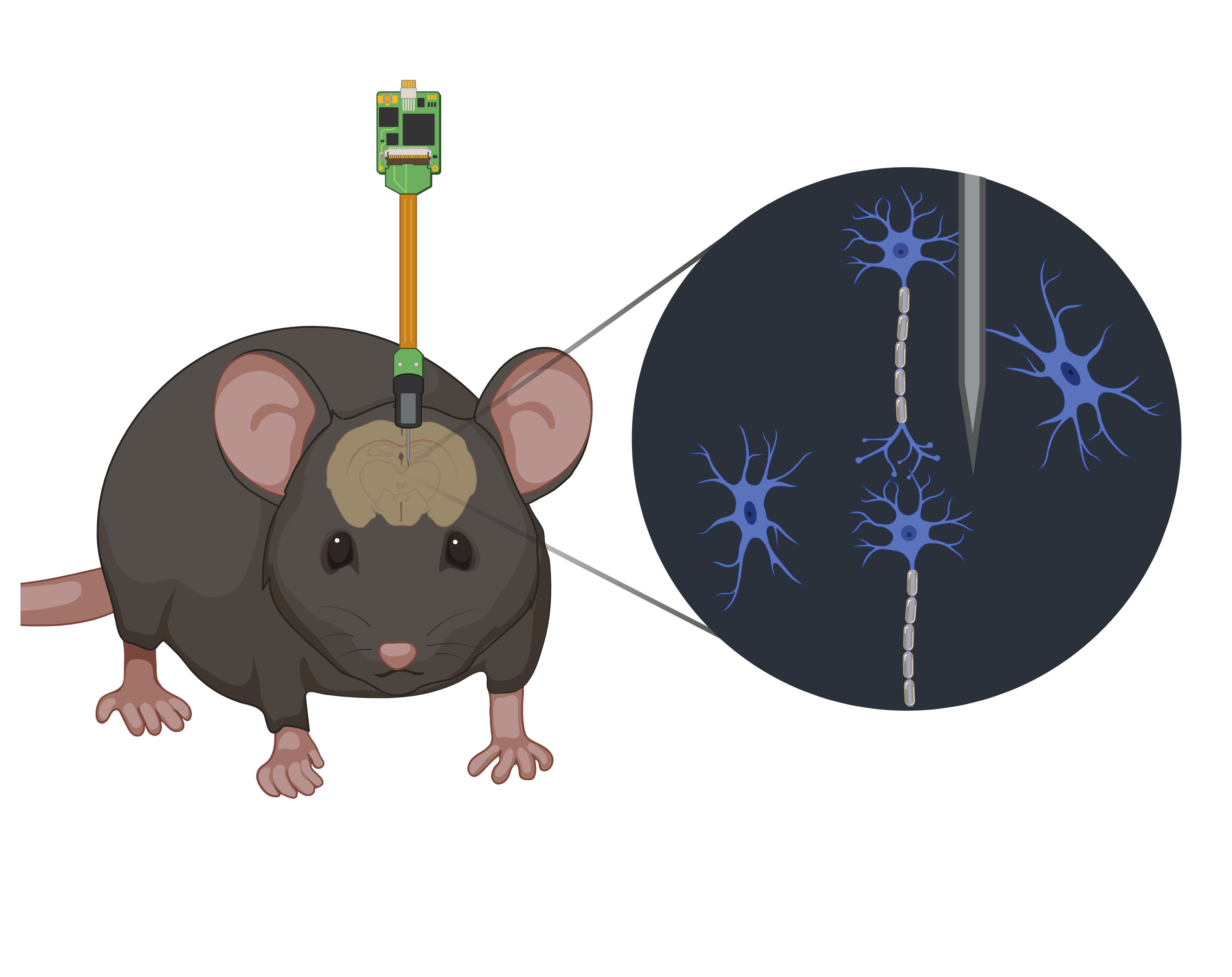
Historically, extracellular microelectrodes have been the primary means to capture neuronal activity. These probes are placed just outside the targeted neuron, allowing it to detect the electrical signals generated by the neuron's activity as changes in voltage, providing insights into the neuron's firing patterns and communication with other neurons. The importance of extracellular microelectrodes lies in their ability to offer a window into the fundamental building blocks of brain function – the individual neurons. However, the capacity to simultaneously monitor a greater number of single neurons is crucial for unraveling coordinated activities. This aspiration led to the development of Neuropixels probes which provide dense, extensive recording sites capable of isolating individual neurons across significant brain regions while minimizing the cross-sectional area to mitigate tissue damage.
The content of this blog post reflects my personal opinions and insights and should not be attributed to my employer or investors. The information provided in this post is for educational purposes only and should not be construed as medical advice. It is crucial to consult with medical professionals for any mental or physical healthcare concerns. All images featured in this blog post were created using Biorender.com under an academic license. These blog posts are derived from excerpts of my PhD thesis, based on research conducted at the University of Oslo, which you can also read on this website.